


Many protozoa live in the human body. Many of them are pathogenic. Our story is about ten of them. The review comes from historical and recent publications.
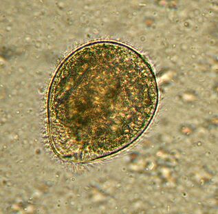
Maximum. BalantidiumBalantidium coli
The largest protozoan is a human parasite and is the only ciliate in the company. Its size is 30 to 150 microns in length and 25 to 120 microns in width. For comparison: the length of the largest malaria parasite is about 15 microns, several times smaller than the swelling of the intestinal cells in which the ureter lives. An elephant in a porcelain shop.
DistributedWhere is the pig-its main carrier. It usually lives in the submucosa of the colon, although it also exists in the lung epithelium in humans. It feeds onEscherichia colibacteria, food particles and fragments of host epithelium. In animals, the infection is asymptomatic. People experience severe diarrhea with bloody mucus secretions (balantidiasis) and sometimes ulcers form in the colon wall. It is rare to die from balance therapy, but it can cause chronic fatigue.
People get infected through dirty water or food containing cysts. The human infection rate does not exceed 1%, and pigs all over the world can be infected.
Treatment with antibiotics, there is no report about the resistance of this ciliate.
was discovered in 1857 by Swedish scientist Malstem. Today, Balantidiasis is associated with poverty and poor sanitation in tropical and subtropical regions.
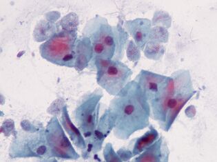
The earliest. Oral AmoebaToothgut Nematode
The first parasitic amoeba found in humans. The description of the amoeba was published in the oldest scientific journal in 1849. Amoeba is found in dental plaque, hence the name gum from the Latin word gum.
Inhabits the mouths of almost all people with sore teeth or sore gums, inhabits gum pockets and dental plaque. It feeds on epithelial cells, white blood cells, microorganisms and red blood cells. It is rare in people with oral health.
This small protozoan, 10–35 µm in size, does not enter the environment and does not form cysts; it can be spread to another host through kissing, dirty dishes, or contaminated food.E. gingivalis (E. gingivalis)is considered the only human parasite, but it is sometimes found in captive cats, dogs, horses and monkeys.
In the early 20th century,E. gingivalis (E. gingivalis)was described as the cause of periodontal disease because it is always present in inflamed tooth cells. However, its pathogenicity has not been confirmed.
The drug that affects this amoebais unknown.
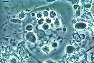
is the most common. Amoeba dysenteryAmoeba
This blood-carrying intestinal parasite penetrates the tissues of the liver, lung, kidney, brain, heart, spleen and genitals. What he can eat: food particles, bacteria, red blood cells, white blood cells and epithelial cells.
Distributionis everywhere, especially in tropical regions. Usually, people get infected by swallowing cysts.
In temperate countries, the amoeba tends to stay in the intestinal lumen and the infection is asymptomatic. In the tropics and subtropics, the pathological process usually begins:E. histolyticaattacks the wall. The reason for the conversion to the pathogenic form is unclear, but several molecular mechanisms that occur have been described. Therefore, it is obvious that the amoeba secretes dissolved substances, breaks through the mucus and kills the cells. Obviously, amoeba can destroy host cells in two ways: by triggering apoptosis or simply by cutting up pieces. For a long time, the first method was considered the only method. By the way, no mechanism for cell suicide with a recording rate of several minutes has been discovered. The second method was described only recently, and the author calls it swallowing syndrome, from the Greek "three" to to. It is worth noting that cell-biting amoeba abandons their prey once they die. Others can swallow dead cells completely. It is assumed that the gene expression patterns of bite and phagocytes are different.
Now, the ability of amoeba to penetrate blood, liver and other organs is related to light swallowing disease.
Amoebiasis is a fatal disease. About 100, 000 people die fromhistolyticainfection every year.
Dysentery amoeba has non-pathogenic twinsdispar, so microscopic examination is not sufficient to diagnose the disease.
To be curedmust be destroyed by movingE. histolyticaand cysts.
DescriptionHemolytic Escherichia coli (E. histolytica), and in 1875 the pathogenicity of its diarrhea patients was determined. The Latin name of amoeba was given by German zoologist Fritz Schaudin in 1903.Histolyticaindicates that the organization is destructive. In 1906, the scientist died of an amoebic intestinal abscess.
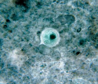
The most common. Small intestineGiardia (G. intestinalis)
Giardia is the most common intestinal parasite and is everywhere. 3-7% of people in developed countries are infected, and 20-30% of people in developing countries are infected. There are about 300 million people.
The parasite lives in the host's duodenum and bile duct, floats in it, works with the flagella, and then attaches to the epithelium with the help of a sticky disc at the bottom of the cell. One million lambs adhere to the epithelium of 1 cm2. They destroy the villi, which interferes with nutrient absorption, causing mucosal inflammation and diarrhea. If the disease affects the bile duct, it is accompanied by jaundice.
Giardiasis is a disease of unclean hands, water and food. The life cycle of a protozoa is simple: there are active forms in the intestines, and stable cysts at the exit of the fecal mass. To be infected, it is enough to swallow a dozen cysts, and these cysts will turn into an active form again in the intestine.
The ubiquitous main secret of LamberiaThe variability of surface proteins. The human body fights the lamb with antibodies, and in principle can develop immunity. But people who live in the same area and drink the same water are infected again and again by the offspring of their own parasites. why? Because during the transition from active phase to cyst (and vice versa), Lamberia will change the antibody-producing protein-variant-specific surface protein. There are about 190 variants of these proteins in the genome, but there is always only one variant on the surface of a single parasite; the rest of the translation is interrupted by the mechanism of RNA interference. This change occurs approximately once every ten generations.
Treat it with antiprotozoal agents with antibacterial activity. The disease disappears within a week, but if the bile duct is infected, it may recur for many years. Cysts are combated by adding iodized water.
was discovered in 1859 by Czech scientist VilémLamblGiardia. Since then, the simplest person has changed a few names, and the current name is known in memory of the discoverer and French parasitologist Alfred Giar, who did not describe LanBia.
Giardia's first sketch was discovered by Anthony van Leeuwenhoek on his own chair. That was in 1681.
By the way, Giardia is also very ancient in evolution, and it comes almost directly from the ancestors of all eukaryotes.
The most intimate. Trichomonas vaginalisTrichomonas vaginalis.
The simplest, is sexually transmitted. It lives in the vagina and men-in the urethra, epididymis, and prostate, through sexual transmission or through wet towels. Babies can be infected through the birth canal.Trypanosoma vaginalishas 4 flagella on the front end and has a short undulating membrane; it releases prosthetic feet when necessary. The maximum size of trichomoniasis is 32 x 12 microns.
Trichomonas spreads more widely than the pathogens that transmit chlamydia, gonorrhea and syphilis. It affects about 10% of women (possibly more) and 1% of men. The latter number is unreliable because it is difficult to detect parasites in men.
Trypanosoma vaginalisfeeds on microorganisms (including the lactic acid bacteria of the vaginal microflora), these microorganisms maintain an acidic environment, so their own optimal pH value is higher than 4. 9.
Trichomonas destroys mucosal cells and causes inflammation. Approximately 15% of infected women complain of symptoms.
Treat with antibacterial drugs. As a precaution, it is recommended to rinse with diluted vinegar regularly.
was described by French bacteriologist Alfred Donne in 1836. The scientist did not know that there was a pathogenic parasite in front of him, but he determined the size, appearance and type of the simplest movement.
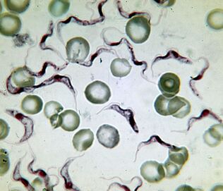
The deadliest. The pathogen of sleeping sicknessTrypanosoma brucei
The cause of African sleeping sickness is the deadliest protozoa. People infected with the virus will die without treatment. Trypanosoma are slender flagella, 15–40 µm long. Two subspecies are indistinguishable in appearance. Disease causedT. brucei gambiensefor 2-4 years.Brucella rosenbergiiis a more virulent, temporary pathogen that dies from it within a few months or weeks.
in AfricaDistributed between the 15th parallel point in southern Africa and the northern hemisphere, within the natural range of the carrier-a blood-sucking insect (collection ofGlossina)Tsetse). Among the 31 species of flies, 11 are dangerous to humans. Sleeping sickness affects the population of 37 countries 9 million kilometers 2 of sub-Saharan Africa2. Up to 20, 000 people get sick every year. There are now about 500, 000 people, of whom 60 million are at risk.
From the fly intestineTrypanosoma bruceienters the human bloodstream, and then enters the cerebrospinal fluid from there and affects the nervous system. The disease begins with fever and inflammation of the lymph nodes, followed by drowsiness, drowsiness, muscle paralysis, fatigue, and irreversible coma.
The lethality of parasites is related to their ability to cross the blood-brain barrier. The molecular mechanism is not fully understood, but it is known that parasites secrete cysteine proteases and use certain host proteins after entering the brain. On the other hand, in the central nervous system, trypanosomes mask immune factors.
The Arab scholar Ibn Halden (1332-1406) left the first description of sleeping sickness in the upper reaches of Niger. At the beginning of the 19th century, Europeans were already aware of the first sign of this disease-swollen lymph nodes on the nape and back (Symptoms of Winterbottom), and slave traders paid special attention to this.
DiscoveryT. bruceiScottish microbiologist David Bruce named after her. He first established trypanosomes in 1903 and collectedThe link between tsetse flies and sleeping sickness.
Treatment methodsDepending on the stage of the disease, medications can cause serious side effects. The parasite has high antigenic variability, so it is impossible to make a vaccine.
The most luxurious. LeishmaniaLeishmania
Leishmania has earned the title of the most extravagant parasites because they live and reproduce in macrophages (cells designed to destroy the parasite).L. donovaniis the most dangerous of them. It can cause visceral leishmaniasis, in layman's terms, hard fever or kala-azar. Almost all patients die without treatment. But the survivors gained long-term immunity.
There are three subspecies of parasites. Mediterranean and Central Asiadonovani infantummainly affects children, usually dogs’ homes.L. donovani donovani(India and Bangladesh) is dangerous for adults and the elderly, and there is no natural reservoir. The United StatesL. donovani chagasi(Central and South America) can live in the blood of dogs.
Lactobacillus donoseli-The length of the flagella does not exceed 6 microns. People are infected after being bitten byPhlebotomusmosquitoes, sometimes through sexual contact, baby-through the birth canal. Once in the blood,L. donovanienters macrophages, which carry parasites through internal organs. Parasites multiply in macrophages and destroy them. The molecular mechanism of survival in macrophages is quite complicated.
Symptoms of the disease-Fever, enlarged liver and spleen, anemia and leukopenia, which are the causes of secondary bacterial infections. Every year, 500, 000 people suffer from visceral leishmaniasis, and about 40, 000 die.
TreatmentSevere illness-intravenous antimony and blood transfusion.
Classification and affiliationDononiellawas determined in 1903 by the famous malaria researcher and Nobel Prize winner Ronald Ross. Its common name is William Leishman (William Leishman), the specific name is Charles Donovan (Charles Donovan), he independently found protozoan cells in the spleens of patients who died of kala-azar at the same time in 1903, One in London and the other in Madras.
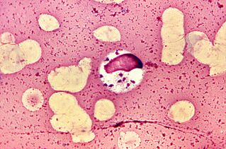
The most difficult life cycle.Babesia
In addition to multi-stage asexual reproduction of sex mites in mammalian red blood cells and intestinal small intestine, Babesia also complicates their development through ovarian transmission. The sporozoites of the protozoa enter the ovary from the small intestine of the female mite and infect the embryo. When the larva of the mite hatches, the Babesia enters the salivary glands and the first bite enters the blood of the vertebrate.
DistributedBabesia in the United States, Europe and Asia. Their natural pools are rodents, dogs and cattle. A person is infected with the following types:B. microti, B. divergens, B. duncaniandvenatorum.
The symptoms of babesiosis are similar to malaria-repeated fever, hemolytic anemia, spleen and liver enlargement. Most people recover spontaneously, but bacillosis is fatal to patients with weakened immune systems.Therapeuticsare still under development, and antibiotics are prescribed, and blood transfusions can be used in severe cases.
DescriptionVictor Babes (1888), a Romanian microbiologist, discovered Bebe insects in sick cattle and sheep. He thinks he is dealing with a disease-causing bacteria calledBoletus. For a long time, Babes disease has been considered an animal pathogen until it was discovered in Yugoslavian sheepdogs in 1957, when the disease died of Bacillus polycephalus infection.
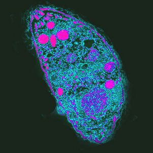
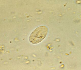
The most influential. The causative factor of toxoplasmosisToxoplasma
Toxoplasma gondiiis the most powerful parasite because it controls the behavior of the intermediate host.
DistributionUneven distribution everywhere. For example, in France, 84% of the population is infected, and in the UK -22%.
infectionsin humans are usually asymptomatic, but weakened immunity can destroy cells in the liver, lung, brain, and retina, causing acute or chronic toxoplasmosis. The infection process depends on the virulence of the strain, the status of the host's immune system and its age-the elderly are not susceptible to infectionT. gondii.
Treating toxoplasmosis with antiprotozoal drugs.
Described in the desert rodents in 1908. The honor belongs to the staff of the Pasteur Institute of Tunisia, Charles Nicolas and Louis Manso.
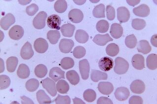
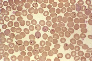
The most pathogenic. Plasmodium plasmodium.
Plasmodium malaria is the most pathogenic parasite in humans. The number of malaria patients can reach 300-500 million, and the death rate during the epidemic is 2 million. The death toll from the disease is still three times that of armed conflict.
Five species of Plasmodium cause malaria in humans:Plasmodium vivax, Plasmodium falciparum, Plasmodium, Plasmodium ovaleandPlasmodium nori, theyIt also affects macaques.
DistributionWithin the vector range-mosquitoesAnophelesrequires a temperature of 16-34°C and a relative humidity of more than 60%.
The deadliest Plasmodium PlasmodiumPlasmodium falciparumThe genome comparison of Plasmodium falciparum and Plasmodium gorilla showed that humans were infected by the ancestors of these monkeys. The emergence of this plasmodium is related to the emergence of agriculture in Africa, leading to an increase in population density and the development of irrigation systems.
The sexual reproduction of Plasmodium occurs in the intestine of mosquitoes. In the human body, it is an intracellular parasite that survives in liver cells and red blood cells and reproduces until the cells rupture. One milliliter of patient’s blood contains 10, 000 to 50, 000 parasites.This disease is manifested by inflammation, repeated fever and anemia. If pregnant, it is dangerous for both the mother and the fetus. Red blood cells infected withPlasmodium falciparumcan block capillaries, and in severe cases, internal organs and tissues may develop ischemia.
Treatmentrequires a combination of multiple drugs and depends on the specific pathogen. Plasmodium is resistant to drugs.